Assami Rosner

and 7 more

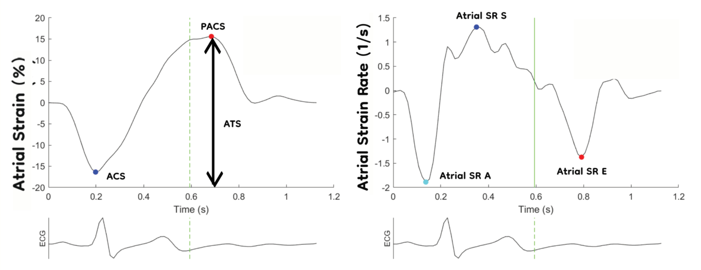
Background Noninvasive assessment of elevated filling pressure in the left ventricle (LV) remains an unresolved problem. Of the many echocardiographic parameters used to evaluate diastolic pressure, the left atrial strain and strain rate (LA S/SR) have shown promise in clinical settings. However, only a few previous studies have evaluated LA S/SR in larger populations. Methods A total of 2033 participants from Norwegian (Tromsø 7) and Russian (Know Your Heart) population studies, equally distributed by age and sex, underwent echocardiography, including atrial and ventricular S/SR and NT-proBNP measurements. Of these, 1069 were identified as healthy (without hypertension, atrial fibrillation (AF), or structural cardiac disease) and were used to define the age- and sex-adjusted normal ranges of LA S/SR. Furthermore, the total study population was divided into groups according to ejection fraction (EF) ≥50%, EF <50%, and AF. In each group, uni- and multiple regression and receiver operating characteristic curve analyses were performed to test LA and LV functional parameters as potential indicators of NT-proBNP levels above 250 ng/ml. Results The mean LA S/SR values in this study were higher than those in previous large studies, whereas the lower references were comparable. In normal hearts, atrial total strain (ATS) and mitral valve E deceleration time (MV DT) were independent factors indicating elevated NT-proBNP levels, whereas in hearts with reduced EFs, the independent indicators were peak atrial contraction strain (PACS) and LV stroke volume. The areas under the curve for these significant indicators to discriminate elevated NT-proBNP levels were 0.639 (95% confidence interval (CI): 0.577-0.701) for normal EF and 0.805 (CI: 0.675-0.935) for reduced EF. Conclusion The results confirm good intrastudy reproducibility, with mean values in the upper range of previous meta-analyses. In the future automated border-detection algorithms may be able to generate highly reproducible normal values. Furthermore, the study showed atrial S/SR as an additional indicator of elevated NT-proBNP levels in the general population, demonstrating the incremental value of both ATS and PACS in addition to conventional and ventricular strain echocardiography. Thus, the LA S/SR may be regarded as an important addition to the multiparametric approach used for evaluating LV filling.

Assami Rösner

and 6 more

Background Left ventricular (LV) systolic and diastolic functions are important cardiovascular risk predictors in patients with hypertension. However, data on segmental, layer-specific strain, and diastolic strain rates in these patients are limited. The aim of this study was to investigate segmental two-dimensional strain rate imaging (SRI)-derived parameters to characterize LV systolic and diastolic function in hypertensive individuals compared with that in normotensive individuals. Methods The study sample comprised 1194 participants from population studies in Arkhangelsk and Novosibirsk, Russia, and 1013 individuals from the Seventh Tromsø Study in Norway. The study population was divided into four subgroups: A. healthy individuals with normal blood pressure (BP), B. individuals on antihypertensive medication with normal BP, C. individuals with systolic BP 140–159 mmHg and/or diastolic BP >90 mm HG, and D. individuals with systolic BP ≥160 mmHg. In addition to conventional echocardiographic parameters, global and segmental layer-specific strains and strain rates in early diastole and atrial contraction (SR E, SR A) were extracted. The strain and SR (S/SR) analysis included only segments without strain curve artifacts. Results With increasing BP, the systolic and diastolic global and segmental S/SR gradually decreased. SR E, a marker of impaired relaxation, showed the most distinctive differences between the groups. In normotensive controls and the three hypertension groups, all segmental parameters displayed apico-basal gradients, with the lowest S/SR in the basal septal and highest in apical segments. Only SR A did not differ between the segmental groups but increased gradually with increasing BP. End-systolic strain showed incremental epi-towards endocardial gradients, irrespective of the study group. Conclusion Arterial hypertension reduces global and segmental systolic and diastolic left ventricular S/SR parameters. Impaired relaxation determined by SR E is the dominant factor of diastolic dysfunction, whereas end-diastolic compliance (by SR A) does not seem to be influenced by different degrees of hypertension. Segmental strain, SR E and SR A provide new insights into the LV cardiomechanics in hypertensive hearts.

Assami Rösner

and 3 more

Background: Decreasing right ventricular (RV) and left ventricular (LV) function after surgical or transcatheter aortic valve replacement (SAVR or TAVR, respectively) is an important risk factor for morbidity and mortality. Although transapical (TA)-TAVR is an independent risk factor for post-procedural mortality, limited knowledge is available regarding long-term changes in RV and LV function. The study aimed to evaluate LV and RV performance following four different AVR procedures, including TA-, transfemoral (TF)-TAVR, and SAVR with and without coronary artery bypass grafting (±CABG). Methods: Patients with severe AS were consecutively included and assigned to TA-TAVR, TF-TAVR, or SAVR ±CABG groups. A total of 130 patients underwent preoperative conventional and strain-rate-imaging echocardiography, with similar controls in the period between 6 and 12 months after the procedure. Results: After AVR, NYHA classes III and IV were reduced from 105 (81%) to 6 (5%) patients. While most of the systolic and diastolic functional parameters indicated improved LV function in the TF-TAVR and both SAVR groups, LV function did not significantly change after TA-TAVR. The right ventricular functional parameters were unchanged or even improved equally after TA-TAVR and TF-TAVR, while they were significantly reduced after SAVR. The Cardiac Index (CI) improved significantly after TF-TAVR from 2.3±0.7 to 2.6±0.7, while staying unchanged after TA-TAVR and SAVR±CABG. Conclusion: This study demonstrated significant changes in LV and RV systolic and diastolic function with functional improvement or deterioration depending on the type of aortic valve replacement. The most significant improvement in CI was observed after TF-TAVR, which is the least invasive procedure.