Wenbo Yang

and 9 more

Background: Hemodialysis (HD) leads to cognitive impairment; however, the pathophysiology of maintenance HD remains unclear. This study aimed to investigate the longitudinal alterations in gray matter volume (GMV) and cerebral blood flow (CBF) in patients on follow-up HD compared with baseline HD, examine the alterations in functional connectivity (FC) by defining co-changed brain regions as seed points, and investigate the correlation between the co-changed brain regions and neuropsychological test scores. Methods: Twenty-seven patients with HD and 30 healthy controls were enrolled in this study. All participants underwent high-resolution T1-weighted imaging, arterial spin labeling, and functional MR imaging to measure GMV, CBF, and FC. The patients on HD were assessed at baseline and 3 years subsequently. Results: The right and left medial superior frontal gyrus (SFGmed.L) exhibited significantly lower GMV and CBF in patients on follow-up HD compared with patients on baseline HD and lower FC between the SFGmed.L and left middle temporal gyrus (MTG.L). Decreased FC between the SFGmed.L and MTG.L was positively correlated with neuropsychological test scores in the follow-up HD group. Conclusions: Reduced GMV and CBF may result in decreased FC between the SFGmed.L and MTG.L, which may be associated with cognitive impairment in patients on maintenance HD. Our findings provide unique insights into the pathological mechanisms of patients on maintenance HD with cognitive impairment.

Mingan Li

and 7 more

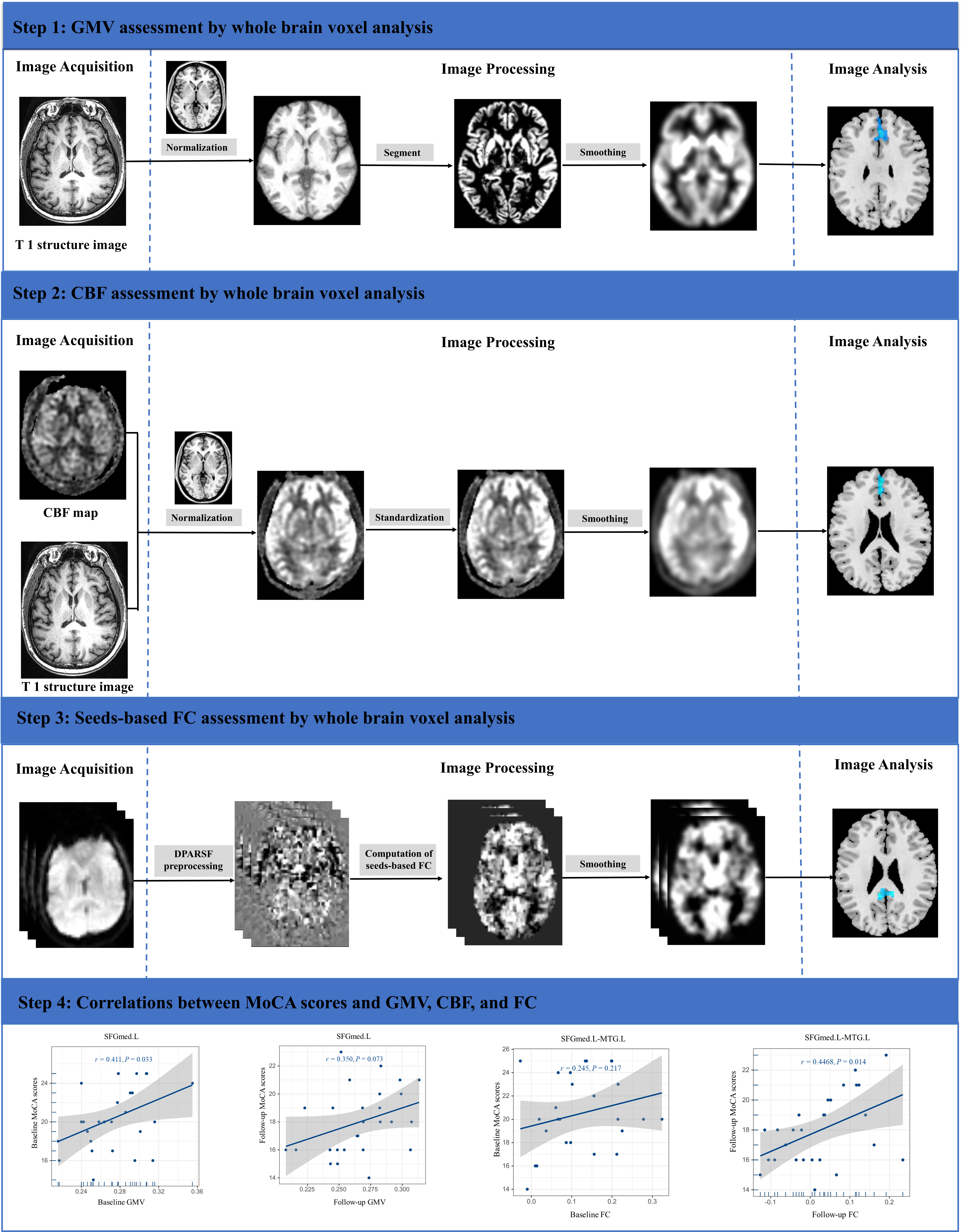
Hemodynamic fluctuations during hemodialysis therapy may result in brain damage, such as white matter hyperintensity (WMH). Cerebral blood flow (CBF) changes occurred before the appearance of WMH. To explore changes in CBF and white matter in hemodialysis patients, patients underwent twice structural and arterial spin-labeling MRI examinations at an interval of three years. Based on the changes in CBF between the baseline and follow-up groups, the hemodialysis patients were divided into two subgroups, increased CBF group and decreased CBF group. Our results showed that patients undergoing hemodialysis exhibited increased cerebral watershed white matter hyperintensities, deep WMH, and periventricular. Among HC, hemodialysis baseline, and follow-up patients, the CBF of gray matter, white matter, and whole matter showed no obvious differences. The CBF of patients with decreased CBF was higher than that of HC at baseline and lower than that of HC at follow-up. Compared with the increased CBF group, obvious development of deep WMH was found in the decreased CBF group for the gray matter, white matter, and whole matter. Therefore, WMH in hemodialysis patients were distributed in the deep white matter, periventricular white matter and cerebral watershed (CW), and progressed with the extension of hemodialysis duration. CBF in hemodialysis patients could manifest as both increased and decreased, and WMH in patients with decreased CBF developed severely with prolongation of hemodialysis duration.