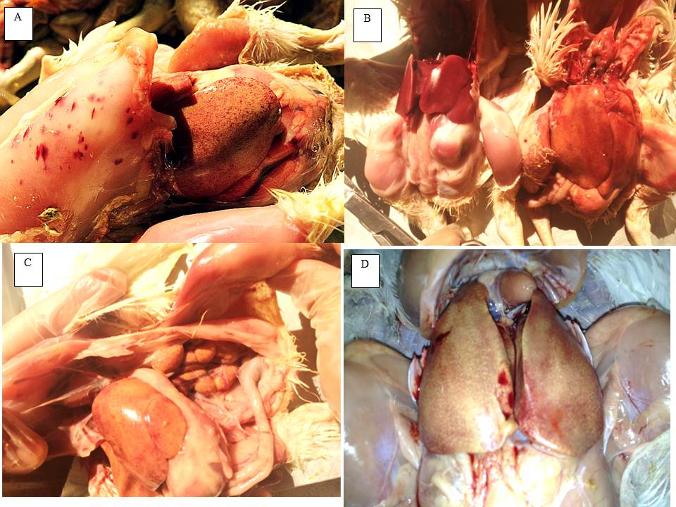
Inclusion body hepatitis is a viral disease caused by Adenovirus group I, and it is worldwide in distribution. The virus is endemic in Sulaymaniyah city, Kurdistan Region of Iraq, and infections occurred in forty-six broiler farms from April 2013 to May 2020. Infected bird’s ages ranged between two days and four weeks. Clinically, birds showed lethargy, huddling with ruffled feathers, inappetence, and yellow, mucoid droppings. Gross lesions included enlarged mottled liver, pale icteric skin, swollen pale kidney, and hemorrhage on the skeletal muscle. Histopathological examinations revealed large intranuclear inclusion bodies in hepatocytes, degeneration and congestion of liver sinusoids, and degeneration of renal tubules, spermatozoa, spermatid, Sertoli cells, and Leydig cells in the testicle. There were intertubular hemorrhages and large vacuoles. The seminiferous tubular lumens were dilated, contained necrotic debris, and were devoid of spermatozoa in the interstitial tissue. Lesions in the testicle are reported for the first time in the present study. RT-PCR was used to detect the virus by amplification of partial 1300 bp hexon genes. The amplified fragments were confirmed by sequencing. Our results concluded that two different genotypes circulate in Kurdistan, and the nucleotide sequence of Kurdistan fowl adenovirus (FAdV) isolates show only 81% homology together. The FAdV/Kurdistan/2013 and FAdV/Kurdistan/2020 belonged to FAdV-E close to USA isolates. On the other hand, the FAdV/Kurdistan/2015 belonged to FAdV-D closer to the Chinese FAdV isolate.